GHK-Cu skin cell research in the laboratory

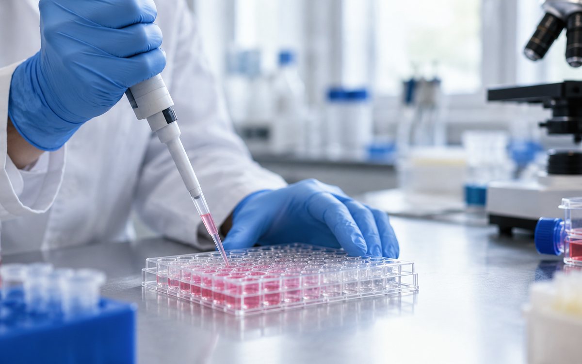
Skin in a culture dish is not a miniature of the human body. It is a controlled system in which the effect of the test substance can be separated from the influence of the medium, cell density or methodology used. That is why GHK-Cu skin cell research significance especially when the question is formulated precisely: does the complex alter the copper-mediated response of fibroblasts, keratinocyte behavior, or extracellular matrix markers in a particular model?
GHK-Cu, also known as glycyl-L-histidyl-L-lysine copper complex, is one of the most frequently studied peptide complexes in the field of skin biology. The interest does not stem from the simple assumption that the substance will have one universal effect. On the contrary, it is a suitable tool for investigating signaling associated with copper homeostasis, cell response to stress, matrix remodeling and gene expression regulation. However, the result depends on the cell model, concentration, exposure time and the form of copper contained in the culture medium itself.
What GHK-Cu represents from a cell biology perspective
GHK is a naturally occurring tripeptide. After binding copper, a GHK-Cu complex is formed, the properties of which cannot be evaluated separately from the availability of the metal ion. Copper is a trace element essential for several enzymes and redox processes, but at inappropriate concentrations it can also change the oxidation state of the cellular environment. Therefore, it is not enough to say in an experiment that the „effects of the peptide“ were tested. It must be clearly defined whether GHK without copper, a pre-prepared GHK-Cu complex or a comparison with a free copper salt is being investigated.
In skin research, GHK-Cu is most often evaluated in the context of dermal fibroblasts, epidermal keratinocytes, and more advanced 3D models. Fibroblasts are relevant for studying the synthesis and organization of the extracellular matrix, including collagen and elastin structures. Keratinocytes, in turn, allow for the monitoring of markers of differentiation, proliferation, migration, and barrier function. Each of these models responds differently to stimuli, so results from one cell line cannot be automatically transferred to another.
GHK-Cu skin cell research: suitable models
For initial screening, primary human dermal fibroblasts or well-characterized fibroblast lines are often practical. Primary cells may better reflect biological variability, but they naturally vary between donors. Cell lines offer greater reproducibility, but may have different metabolic activity and response compared to the primary tissue.
Keratinocyte models are suitable if the hypothesis is focused on the epidermis. In their culture, particular attention should be paid to the composition of the medium, calcium concentration, and differentiation status. These variables can significantly affect the expression of keratins, filaggrin, involucrin, or other markers, thereby overlapping or mimicking the response to the test compound.
If the research question involves communication between the epidermis and dermis, a co-culture or reconstituted 3D skin model may be a more valuable option. Such systems provide a more biologically relevant context, but are more challenging to standardize, image, and interpret. For many laboratories, it makes sense to proceed from 2D screening to 3D validation once a consistent signal is obtained.
Which parameters make sense to monitor?
Cell viability alone is a useful starting point, but does not provide an answer to the mechanism. Metabolic assays may respond to changes in redox activity and therefore cannot be interpreted as direct evidence of cell number in the case of copper complexes. They should be combined with cell counting, morphological assessment, or membrane integrity analysis.
In fibroblasts, the expression of genes and proteins associated with the extracellular matrix can be monitored, for example collagens, fibronectin, elastin, matrix metalloproteinases and their inhibitors. The difference between the change in expression and the actual deposited matrix is important. Quantitative PCR, immunofluorescence, Western blot or methods assessing collagen deposition answer different parts of the same biological question.
In keratinocytes, proliferation, migration in gap closure models, markers of differentiation, and transcriptional markers of barrier function may be relevant. However, the scratch assay model requires caution. Gap closure may be a consequence of migration, proliferation, or a combination of both processes. Without appropriately designed controls, these mechanisms cannot be reliably separated.
Concentration, time and control groups make the difference
With GHK-Cu, it is sensible to work with a concentration range, not a single point. Nonlinear response is not uncommon with bioactive complexes: the concentration at which a measurable change in the selected marker occurs may not be appropriate for a different endpoint or longer exposure. A pilot experiment should verify cell tolerance, solubility, and solution stability before a larger series is initiated.
The design is based on at least an untreated control and a vehicle control. If the contribution of copper is being investigated, a comparison with GHK without copper and an equimolar free copper control may also be methodologically valuable. Such a design helps to distinguish whether the observed phenomenon is related to the peptide moiety, copper complexation, or a general effect of the copper ion in the given medium.
Serum conditions must also be included in the interpretation. Serum components can bind both peptides and metals, alter their availability to cells, and influence the growth profile of the culture. Therefore, an experiment in serum-containing media and an experiment in serum-free conditions may not yield the same result. Neither approach is automatically better – the choice must be based on the biological question and model validation.
Analytical sample quality is not a formality
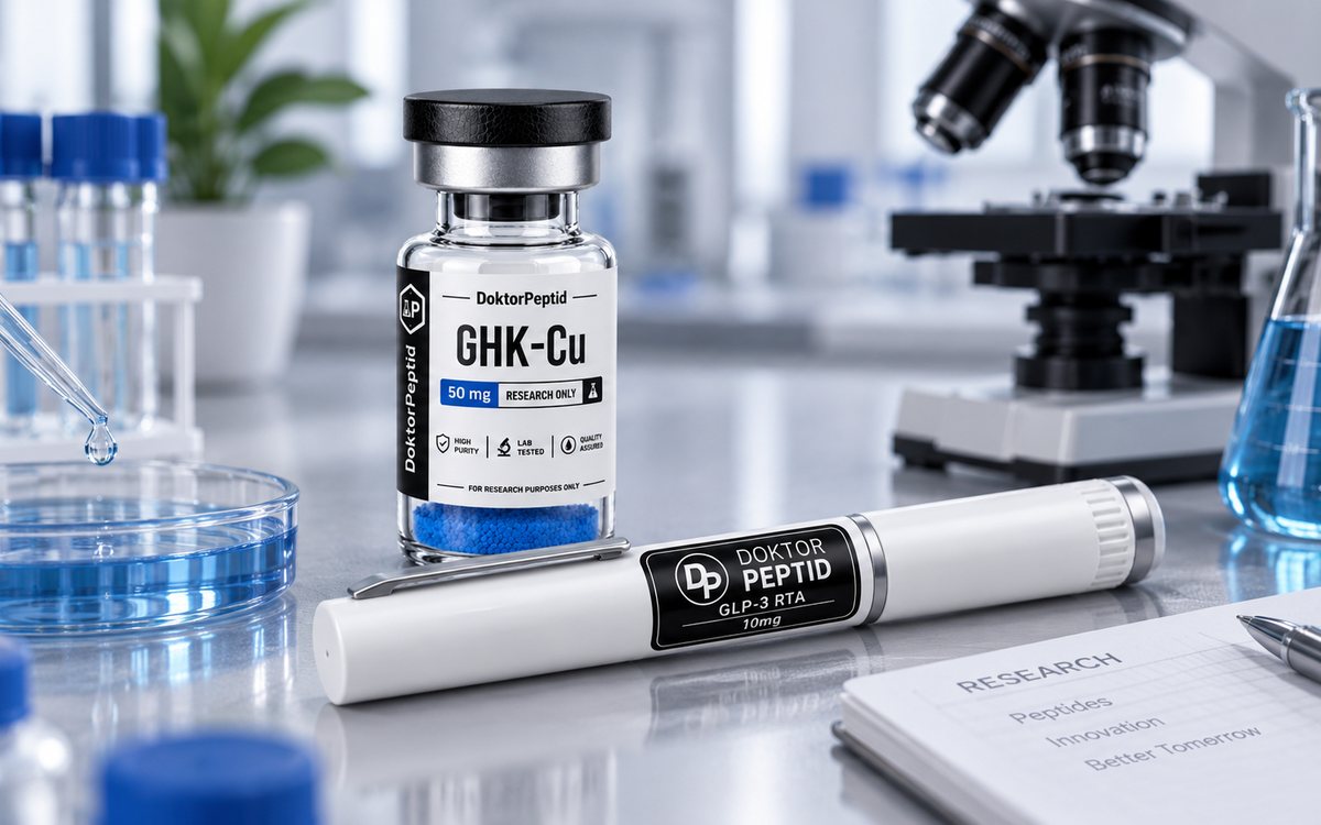
In cell-based assays, an impurity, an inappropriate counterion, residual solvent, or a difference between batches may cause an interpretation that is incorrectly attributed to GHK-Cu. It is therefore advisable to work with a research compound declared purity at least 99 % and request batch documentation. HPLC analysis provides an important indication of chromatographic purity, while mass spectrometry supports verification of the identity of the molecule.
Quality control also has a practical impact on reproducibility. If a laboratory changes a batch, changes suppliers, or compares results between sites, the analytical protocol and accurate material records are part of the experiment, not extra administration. DoktorPeptid states a declared purity of ≥99 % for research materials and provides the option to request an analysis protocol for a specific batch, which simplifies the documentation of input material.
Preparation and storage before the experiment
The working solution should be prepared according to the internal laboratory procedure and taking into account the compatibility of the solvent with the cell system. At low concentrations tested, even a small difference in the volume of vehicle can affect the result. Therefore, keep the vehicle the same in all relevant groups.
With peptide materials, it is important to limit repeated freeze and thaw cycles. Aliquoting after reconstitution reduces the risk of both degradation and contamination. Protection from light, appropriate temperature, and a clear record of the date of preparation are simple steps that increase the chance that the difference between wells will be biological, not technical.
How to read the results without jumping to conclusions
A positive change in one marker does not demonstrate a complex effect on the skin. Increased expression of a particular gene may not mean a functional change in the tissue and the result from a 2D monoculture is not clinical evidence. Likewise, a negative result may not automatically mean that GHK-Cu has no biological activity in the model – it may be an inappropriately chosen endpoint, time point or culture conditions.
The most valuable data is generated when an observed phenomenon is confirmed by an independent method, in biological replicates, and with appropriate statistical analysis. Transparently record cell passage, seeding density, medium composition, concentration, exposure time, material batch, and sample exclusion criteria. GHK-Cu skin cell research set up in this way yields results that are usable for further hypotheses, not just attractive graphs.
GHK-Cu is intended for in vitro scientific purposes only. It is not a drug, food supplement, or material for human or animal consumption. With a well-defined model, analytically validated material, and disciplined controls, it can be a useful tool to ask more precise questions about skin cell biology.
Related products
Produkty, ktorých sa táto téma týka: GHK-Cu – skladom na Slovensku, Baktériostatická voda – skladom na Slovensku. Ceny a dostupnosť nájdete v sekcii všetky produkty, doplnky ako dávkovacie perá a zvýhodnené balíčky sú v samostatných kategóriách.
Všetky produkty sú určené výhradne na laboratórny výskum.