
Can research peptides affect cell viability?
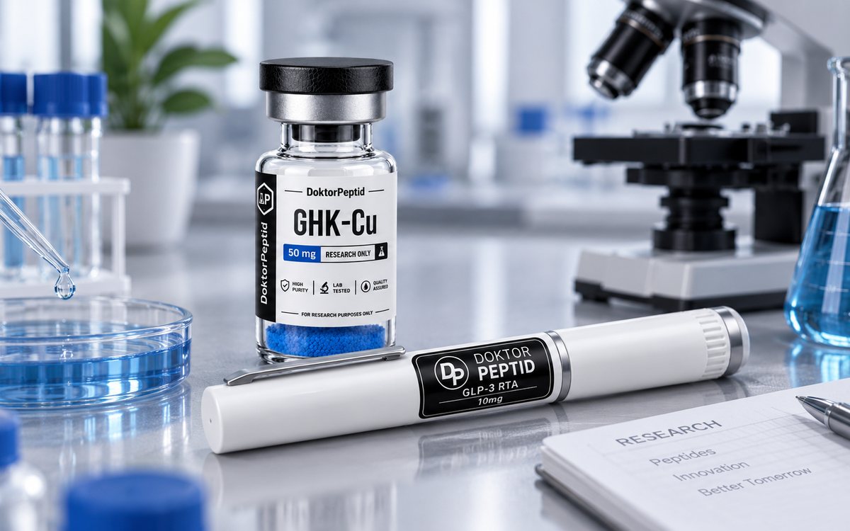
Cell viability is not a simple answer of „the cell is alive“ or „the cell is not alive.“ In the laboratory, the same intervention can alter metabolic activity, division rate, membrane integrity, and apoptotic signaling—and each of these phenomena is captured by a different test. The question of whether can research peptides affect cell viability, therefore, requires a precisely defined model, an appropriately chosen method, and a disciplined interpretation of the results.
Peptides are of interest in cellular research mainly because of their ability to interact with receptors, membranes, enzymes or signaling pathways. However, this does not in itself mean that a particular compound „helps“ or „harms“ cells. The outcome depends on the cell type, concentration, length of exposure, composition of the medium, culture density and analytical readout. In in vitro research, it is crucial to replace general expectations with a measurable hypothesis.
What is actually measured by cell viability?
The term viability is often used as an umbrella term, but individual tests monitor different biological parameters. A metabolic test may indicate a change in mitochondrial activity without directly changing cell number. Conversely, a membrane integrity test will detect damage or lysis, but may not detect early apoptosis or slowing of proliferation.
When evaluating the effect of an investigational peptide, it is therefore useful to determine in advance which question the experiment is intended to answer. Is it acute cytotoxicity? Is it a change in proliferation after 24 to 72 hours? Is it the ability of cells to recover from a defined stressor? Or is it a shift in apoptotic signaling? One plate and one colorimetric assay are usually not sufficient for a reliable biological conclusion.
Metabolic activity is not the number of living cells
Tetrazolium salt reduction assays, such as MTT, XTT or WST, and fluorescent methods with resazurin are often used because they are practical for multi-chamber formats. However, their signal depends on the enzymatic and metabolic activity of the cells. If the peptide affects redox metabolism, mitochondria or proliferation, the result may change even without direct cell death.
Similarly, ATP testing provides a sensitive indirect image of active cells, but ATP is a dynamic marker. A lower signal may be related to a decrease in cell number, temporary metabolic adaptation, or technical variability in lysis. Therefore, results should be interpreted precisely: for example, as a change in metabolic signal relative to control, not automatically as evidence of cytotoxicity.
Membrane, apoptosis and proliferation complete the picture
Membrane damage can be verified by measuring LDH release or by staining live and dead cells. Annexin V in combination with propidium iodide or a similar stain helps to distinguish early apoptotic, late apoptotic, and membrane-damaged populations. Direct cell counting, confluence image analysis, or nucleoside analog incorporation assays are useful for assessing cell growth.
Combining at least two orthogonal methods significantly reduces the risk that an experiment confuses a metabolic effect with an effect on viability. For example, if the ATP signal decreases but cell number, membrane integrity, and apoptotic markers remain unchanged, interpretation should remain cautious.
How research peptides can affect cell viability
The effect of a peptide can arise through multiple mechanisms. Some research compounds are studied in the context of signaling associated with cellular stress response, migration, inflammation, energy metabolism, or differentiation. Others may elicit an undesired response at inappropriate concentration, insufficient solubility, or when interacting with a specific cell model.
For peptides being studied in the field of regeneration, such as BPC-157 or TB-500, it may be interesting to monitor viability in addition to cell migration and markers of damage response. In skin or fibroblast models, it may GHK-Cu require separate assessment of the influence of the peptide component and copper concentration. For compounds investigated in neurobiology, such as Selank or Semax, the relevant readout depends on the type of neuronal or glial cell and the stress model chosen.
These examples do not constitute therapeutic claims or predictions of outcome. Their significance is to illustrate the need to align the peptide, the cell model, and the biological question. An effect observed in one line cannot be extrapolated to another line, tissue, or organism without additional data.
Concentration, timing, and formulation often matter more than expectation.
A concentration curve is the foundation of a quality experiment. Testing a single concentration can create a misleading picture, especially with a nonlinear response. It is more practical to prepare a logarithmic range of concentrations based on pilot data, compound solubility, and cell model properties. Concentrations that cause precipitation or excessive vehicle in the medium should be avoided.
The exposure time is also important. An acute 4- or 6-hour experiment can capture the early stress response, while a 24-, 48-, or 72-hour measurement will better demonstrate effects on proliferation and persistence of the cell population. For longer incubations, peptide stability in culture medium, degradation by proteases, medium exchange, and possible adsorption to plastic surfaces must be considered.
Formulation and handling are equally important. Inappropriate pH, repeated freeze-thaw cycles, microbial contamination, or improperly prepared vehicle can affect cells regardless of the properties of the peptide itself. Therefore, each series of experiments should have a vehicle control and clearly documented reconstitution, dilution, and storage procedures.
Controls that distinguish signal from artifact
A quality experiment does not only consist of a test sample and a negative control. When measuring viability, it is advisable to include a positive damage control to confirm that the assay used can capture the expected signal drop in the given system. Empty wells without cells help to detect substance interference with the reagent or optical measurement.
Some peptides, excipients, or formulation components may affect absorbance, fluorescence, or luminescence. If a sample produces a signal even in a well without cells, the metabolic assay result cannot be interpreted without correction. This step is particularly important for colored or redox-active materials.
Replicates have their own role. Technical replicates reduce the impact of pipetting variability, biological replicates test repeatability across independent experiments. For cell lines, it is also appropriate to monitor passage, morphology, confluence at seeding, and regularly check for mycoplasma contamination. Without these data, even a statistically significant difference may be biologically uncertain.
Why the analytical quality of a research peptide is part of study design
If a subtle shift in viability is examined, identity and material purity are not an administrative detail. Impurities, degradation products, synthesis residues or differences between batches can alter results, especially in sensitive cell models. Comparing data between experiments only makes sense when the material is thoroughly characterized.
When procuring research peptides, it is therefore appropriate to require a declared purity of at least 99 %, HPLC analysis and, if necessary, confirmation of identity by mass spectrometry. A batch-specific analysis protocol allows the laboratory to link the results obtained to the material used. Equally important are clear storage instructions, batch traceability, and product protection during transport.
DoktorPeptid approaches these parameters as a practical part of research reproducibility: for research material, documentation has the same value as a correctly adjusted pipette or a validated protocol. All products are intended exclusively for in vitro scientific purposes, not for human or veterinary use.
How to formulate the result without unnecessary simplification
A precise result is more useful than a broad statement. Rather than stating that the peptide „improved viability,“ it is methodologically more correct to state the cell model, concentration, duration of exposure, assay used, effect size, and comparative control. If the change was confirmed by multiple methods, this should be clearly stated. If it was not confirmed, this result is also relevant information for further study design.
The best peptide experiments don't start with the question of what the material should do. They start with the question of what biological phenomenon we want to reliably measure—and whether we have controls, an analytically validated batch, and methods that can distinguish a real effect from a laboratory artifact.
Related products
Produkty, ktorých sa táto téma týka: BPC-157 – skladom na Slovensku, TB-500 – skladom na Slovensku, GHK-Cu – skladom na Slovensku, Semax – skladom na Slovensku. Ceny a dostupnosť nájdete v sekcii všetky produkty, doplnky ako dávkovacie perá a zvýhodnené balíčky sú v samostatných kategóriách.
Všetky produkty sú určené výhradne na laboratórny výskum.